
Linda Moy, MD, is professor and vice chair for artificial intelligence in the Department of Radiology at NYU Grossman School of Medicine and clinician-researcher with the Center for Biomedical Imaging and the Center for Advanced Imaging Innovation and Research at NYU Langone Health. She is an internationally recognized expert on breast imaging, editor emerita of Radiology, the flagship scientific journal of the Radiological Society of North America, and member of the American College of Radiology Commission on Breast Imaging. She is known as an advocate for breast cancer screening standards, investigator of machine learning applications in breast imaging, thought leader on the benefits and risks of artificial intelligence in radiology, and prominent voice in the field’s emerging conversation on environmental sustainability. In May, Dr. Moy became the 2026-2027 vice president–elect of the International Society for Magnetic Resonance in Medicine, the world’s largest professional organization dedicated to the advancement of MRI, with a membership of more than 8,000 radiologists, technologists, imaging scientists, and medical device engineers. Our conversation was edited for clarity and length.
You are known for your work on breast cancer screening, breast imaging research, and AI applications in radiology. But looking way back, what brought you to breast imaging in the first place?
I was drawn to radiology because I liked novel technology and was fascinated by the idea that you could look inside the body and figure out what was wrong—like a detective solving a puzzle. During residency, however, I realized that radiologists often work behind the scenes. We perform detailed image analyses and provide recommendations, but it is usually the referring physician who communicates the findings to the patient, discusses management, and guides the next steps. And I realized that I wanted to work with a patient population where I could make a meaningful difference, help shape clinical decisions, and build relationships with patients. That’s what led me to breast imaging.
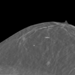
The foundation of breast imaging is screening mammography. In the U.S. most women begin annual screening at age 40, and those at higher risk may require additional imaging tests. What I find especially rewarding is the opportunity to interact directly with patients—to explain their results, answer questions, and help them understand the path forward. Being able to combine advanced imaging technology with compassionate patient care is what has made breast imaging such a fulfilling career for me.
And efforts to bring radiologists out of the shadows, so to speak, accelerated in recent years. At NYU Langone, radiologists participate in virtual rounds and create lay-friendly video reports. Can you talk about that evolution of the radiologist’s role?
Radiologists typically think of the report as the product, but our reports are usually very technical and filled with jargon. We realized that in order for the referring physicians and patients to better understand what the next steps are and to make sure that patients come back for the follow-ups, we needed to communicate better.
This is where I think AI has made a big difference. It is now much easier to translate complex medical information into patient-friendly materials. In breast imaging, we are already required by law to provide patients with understandable summaries of their results, but new technologies allow us to go much further. We can create reports in multiple languages, tailor explanations to different reading levels, generate video summaries, and develop educational content that explains procedures such as biopsies and what patients can expect throughout the process.
By improving communication and making imaging more accessible and understandable, we’re becoming more visible members of the healthcare team and more active partners in patient care.
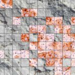
Through research with colleagues at NYU Langone, you have contributed to advancing AI applications for breast imaging; and you have coauthored many original research articles, reviews, meta-analyses, guidelines, and editorial opinions on AI in radiology—including in mammography, digital breast tomosynthesis, breast ultrasound, MRI, and PET/MR. One of the things you have looked at are large language models. Can you talk a about the balance of opportunity versus risk in LLMs?
Large language models are an AI technology trained on an enormous amount of text to predict the next word in a sentence, and they’ve become excellent summarization tools. Every healthcare provider needs help communicating and documenting their encounter with a patient. With LLMs, we can use summarization tools to almost instantaneously provide transcribed reports. We see this now at NYU Langone beyond radiology, with physicians asking patients’ permission to record the visits and an AI automatically generating the reports. But the devil is in the details.
Most of the time the summary is clear and consistent with the conversation. However, the AI is only as good as the data it was trained on, so some biases will appear. I’ll give an example: the patient may say ‘I have a non-productive cough and am a bit short of breath.’ Maybe the text that the LLM trained on had the word ‘fever’ frequently appearing in the vicinity of ‘cough’ and ‘shortness of breath.’ This may lead the LLM to add ‘fever’ to the transcription. If you’re not careful double-checking that, you may interpret the report as listing three common signs of pneumonia and start the patient on a course of antibiotics. That’s just a subtle one-word mistake, but it’s an example of an unintended consequence. Overall, to have that report—provided that a human reviews it—is really a game-changer in terms of freeing up a doctor’s time to do other tasks.
LLMs are excellent tools, but currently, they cannot run autonomously in the background without a human checking them.
A decade ago, when AI was breaking through to public awareness, Geoffrey Hinton famously predicted that radiologists would soon become obsolete. It’s become popular to point out that the prediction hasn’t come to pass. Even so, machine learning has affected every step in the imaging process, from data acquisition to image formation to interpretation to reports. In your view, how has the technology affected the profession so far and what is its potential at this point?
Let me shift to AI as a computer-vision tool—that is the context that Geoff Hinton had in mind. He was focused on the one task of looking at a particular study and generating a specific prediction, for example: fracture or no fracture. Where radiologists provide the greatest value is in bringing together the full clinical picture. We consider why a patient is undergoing an examination, review the electronic health record, compare prior imaging studies, and integrate all of this information to deliver a more nuanced interpretation and guide next steps.
Image interpretation is only one component of the imaging pathway. Radiologists are also involved in scheduling and protocoling examinations, advancing imaging technology, communicating results to referring physicians and patients, and participating in multidisciplinary conferences to help manage complex cases. These contributions are difficult to quantify and cannot be reduced to the isolated task of reading an image.
AI is not panacea. Regulatory approval is often based on retrospective studies that show high diagnostic accuracy, but implementation in clinical practice is different, because we don’t know in advance how humans will interact with the AI system, how much they’ll trust it, or how it will perform in a population it’s never seen. Radiologists have been using AI to detect breast cancer for years, and we’re all realizing that the hard part is how to make AI work in our individual workflows, which may vary even among practices in a single healthcare system.
You are the inaugural vice-chair for artificial intelligence in NYU Langone’s radiology department. What kinds of projects and goals do you have in this role?
The credit for the vision and the drive here goes to [NYU Langone’s radiology department chair] Dr. Recht—he has built on very successful research collaborations with Visage and Siemens. We have successfully made our exams faster and shorter while maintaining great image quality to address issues of access. Also, we understand that if you have an AI system, it has to be interoperable. In other words, it has to be easy for the radiologist to use, to see the results, and to incorporate them with other information. We’ve looked at a number of AI systems and evaluated whether they’d be a good fit for our department, as well as developed home-grown AI systems. Currently, the hottest topic is image-to-text analysis.
Can you describe what image-to-text analysis is?
The concept is based on training models to learn associations between images and words, initially done for image captioning. In radiology image-to-text uses AI to interpret medical images and automatically generate diagnostic findings or complete reports. These systems are trained on large datasets of paired medical images and radiologist reports and learn to translate visual information into clinical language.
The technology combines two components: a vision language model, or VLM, that analyzes images or videos and identifies relevant findings; and a large language model, LLM, that converts those findings into coherent, clinically meaningful text. These multimodal AI systems can assist with report generation and streamline radiology workflows.
In recent years, you have taken up the subject of sustainability, and at the 2025 meeting of the European Congress of Radiology you advocated for reducing radiology’s impact on marine ecosystems. Some of the profession’s impacts include packaging waste, energy consumption, and the use of gadolinium—an element in contrast agents that doesn’t break down and ends up in the oceans. How did you come to view the field in this very broad context?
After Covid there was a new awareness of healthcare disparities and an awakening that we’re not a silo. It also became apparent that radiology as a field contributes a lot to the greenhouse gas emissions worldwide, and we had not focused on that lens before.
We had thought that if we could perform imaging exams quickly, produce high-quality images, help patients receive an accurate diagnosis, and ultimately improve outcomes, then we’ve done our job well. More recently, we have begun asking: Can we continue to do that while also minimizing the environmental impact?
This movement gained early momentum in Europe and Asia, where sustainability has become a major focus, and has been embraced by a younger generation of radiologists who are eager to address these challenges.
Sustainability is an area that requires collaboration across the entire imaging ecosystem. Pharmaceutical companies can develop more environmentally friendly contrast agents. Scanner manufacturers can design hardware and software that consume less energy and incorporate reusable or repurposed components. Radiology departments can reduce waste, improve operational efficiency, and lower energy consumption while maintaining high-quality patient care. But the first big step is a collective commitment to engage in the sustainability movement.
You have just become the 2026-2027 vice president–elect of the International Society for Magnetic Resonance in Medicine. This is a four-year track. After serving as vice president–elect, you will go on to consecutive one-year terms as the society’s vice president, president, and past president. What does this responsibility mean to you?
I was privileged to have been nominated for this leadership role by the committee and the board of trustees. After the nomination, the entire membership votes, so it’s a real honor that they picked me.
This is the largest MRI organization in the world. The members include radiologists and technologists, MR researchers, engineers, physicists, chemists, data scientists, AI researchers, imaging device manufacturers, pharmaceutical companies, and a large number of trainees. This diversity is one of the society’s greatest strengths. We come together to form a vibrant community where people are thinking about ways to use MRI to address some of the hard questions in medicine. For example, can we detect Alzheimer’s disease earlier, before significant cognitive decline occurs, and identify opportunities for intervention that may slow or prevent progression?
The ISMRM annual meetings bring together thousands of professionals from across the globe to share scientific discoveries, technological innovations, and advances in clinical practice. We rotate sites for the annual meeting between North America, Europe, and Asia, and this year was the first time we held the annual meeting in Africa. It’s exciting because the broad range of stakeholders creates opportunities for collaboration and innovation that wouldn’t be possible within a single discipline.
Do you remember your first ISMRM?
It was in 2006 in Seattle. Back then, we still had the traditional poster sessions—thousands of posters lined up in row after row. I remember being very nervous as someone who was very famous approached my poster and said, ‘Can you tell me about your research please?’ I probably stumbled through my answer, but that brief interaction turned out to be important. Later in the meeting, I recognized him, said hello, and that simple connection was the first step in what became a long-term mentorship.
That experience captures what makes the ISMRM so special.
As we talk, I’m getting a sense of curiosity and optimism from you. What would you say to that?
I think this is an exciting moment for our field. Radiology will continue to play a central role in patient care, and the demand for imaging is only going to increase as the population ages. At the same time, challenges related to access, workforce capacity, and healthcare costs will become more pronounced. This is a great time for radiologists to use AI and other emerging technologies to work smarter and deliver greater value. Given our finite resources, how can we provide the right test, at the right time, for the right patient?
What excites me most is that imaging is beginning to move beyond disease detection and treatment monitoring into the realm of prevention. Advances in AI, foundation models, and opportunistic screening are allowing us to identify signs of disease before symptoms develop. This shift toward detecting subclinical disease has the potential to fundamentally change how we care for patients.
That’s where research centers like our Center for Biomedical Imaging and the Center for Advanced Imaging Innovation and Research at NYU Langone are well positioned to lead this transformation. We can rethink how images are acquired, develop imaging biomarkers that provide deeper insight into disease biology, and create innovative solutions to some of healthcare’s most pressing challenges.
So yes, I am optimistic—but it is an optimism grounded in the recognition that there is still a great deal of work to do. I’m really excited for what will happen for our field in the next five to ten years.
Related Stories

Congratulations to Linda Moy on becoming the 2026–2027 vice president–elect of the International Society for Magnetic Resonance in Medicine.

The fastMRI dataset now includes curated breast MRI data to boost AI innovation in radial, dynamic contrast-enhanced, ultrafast MRI of the breast.
Recent research shows that sodium MRI can predict cancer response to chemotherapy. The National Cancer Institute is funding NYU Grossman School of Medicine to develop and validate the method.
NYU Langone researchers won a deep learning challenge to detect lesions in digital breast tomosynthesis (DBT) images. We discuss the obstacles that DBT poses to deep learning and look at how our team navigated them.