The National Cancer Institute has awarded $3.5 million over five years to researchers at NYU Grossman School of Medicine to develop a multinuclear MRI technique for monitoring breast cancer response to neoadjuvant chemotherapy, also known as preoperative or primary systemic therapy.
The NIH-funded project has the potential to fill an unmet need in breast cancer treatments that begin with chemotherapy with the goal of shrinking large or aggressive tumors in order to enable mastectomy or promote breast-conserving surgery. Neoadjuvant chemotherapy doesn’t succeed in all patients but there are no reliable early-monitoring tools to determine whose cancer is responding and whose isn’t before several treatment cycles. Ryan Brown, PhD, and Guillaume Madelin, PhD, associate professors at NYU Grossman School of Medicine and scientists at the Center for Advanced Imaging Innovation and Research, propose to develop an MRI method that can tell the difference.
“Anatomical and structural changes may not be observed in MRI images for months,” said Dr. Brown, referring to conventional MRI, which senses signal from the body’s hydrogen atoms in order to provide detailed images of organs and tissues. “That causes non-responders to be missed.”
In the new project, the research team is aiming to develop an MRI technique that also tunes into signal from sodium atoms. The goal of combining sodium and hydrogen (in MRI parlance, proton) data is to enrich images with metrics of the tumor’s viability.
“The advantage of sodium MRI is that it’s related to metabolic information, rather than structural,” said Dr. Brown. “In a cell you have a sodium-potassium pump. That pump is responsible for removing sodium ions and bringing in potassium ions. … If chemotherapy is working properly and making the [cancer] cell not viable, then we expect to see a change in the sodium level.”
The researchers predict that metabolic damage to cancer will be measurable long before structural degradation becomes visible. “We believe that we’ll be able to predict patient response within one or two weeks of the first cycle of chemotherapy,” said Dr. Brown.
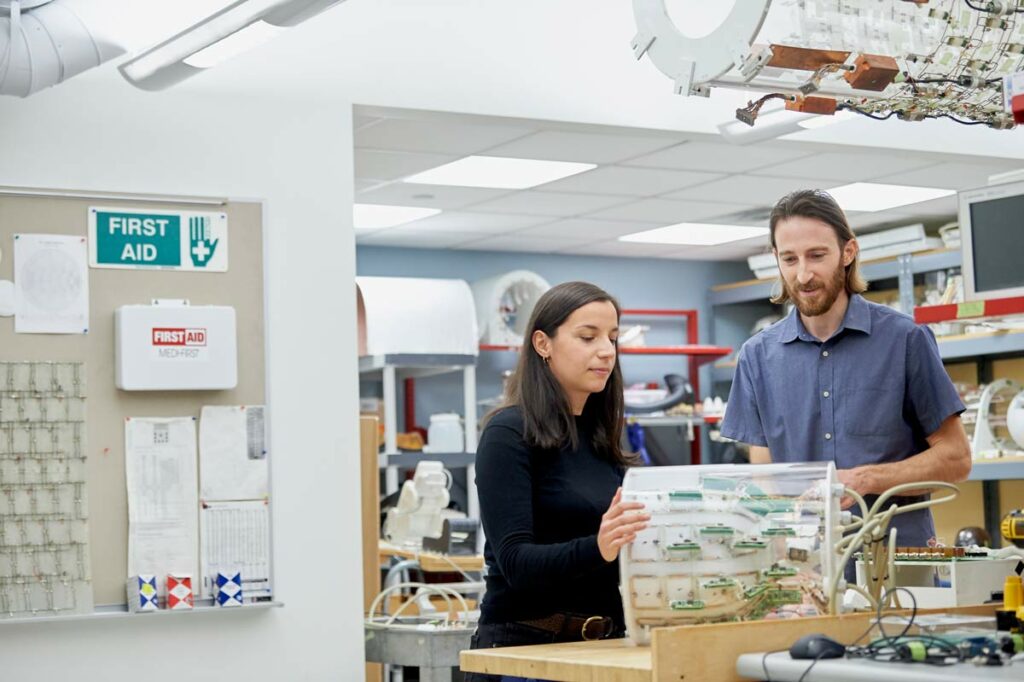
The primary challenge of sensing sodium signal is that it’s between 3,000 and 20,000 times weaker than hydrogen signal, leading to noisier images and longer scan times—hurdles that keep multinuclear MRI in the lab and out of the clinic. However, recent work led at the Center for Advanced Imaging Innovation and Research by Carlotta Ianniello and Zidan Yu, then PhD candidates in Grossman School of Medicine’s biomedical imaging and technology program, has laid a foundation for overcoming these barriers. Dr. Ianniello’s research involved the development of a dual-tuned sodium-proton MRI coil for breast imaging; Dr. Yu’s, the development of a pulse sequence to simultaneously acquire quantitative sodium and proton data.

Separately, a group of imaging scientists led by Olgica Zaric, PhD, in Vienna, Austria, and Erlangen, Germany, showed in a recent feasibility study that sodium MRI can predict whether patients would respond to neoadjuvant chemotherapy after just one treatment cycle—a finding that Drs. Brown and Madelin used to bolster their argument to the National Cancer Institute. “That definitely gave us confidence that our intuition was correct,” said Dr. Brown.
“The Vienna group … measured only total sodium concentration, whereas we plan to measure both intracellular sodium concentration and volume fraction along with sodium relaxation times,” using a technique NYU Langone’s team has already developed, said Dr. Brown. “We expect these measures to provide more information about cell membrane integrity, ion transporters, and loss of homeostasis.”
The proof-of-concept research performed independently at NYU Langone and in Europe was in each case done at 7 Tesla, a high magnetic field strength approved for clinical use only five years ago, currently limited to a single MRI scanner model, and still rare in imaging clinics—even in highly developed healthcare markets like New York City.
Drs. Brown and Madelin propose to develop the multinuclear method for 3 Tesla, a lower but more commonly available field strength, and to combine sodium measurements with more conventional MRI data in order to form an informative snapshot of a tumor’s microenvironment. Another factor motivating the shift to 3 T has been lack of compatibility between 7 T scanners and a type of mediport widely used at NYU Langone (mediports are implanted catheters that facilitate intravenous drug delivery). Although 3-Tesla MRI scanners are more prevalent in radiology clinics, lower field strength means less signal and more noise.
“One of the new challenges will be to overcome the signal-to-noise ratio deficit, and we hope to do that by developing a custom coil and a more efficient pulse sequence sampling scheme,” said Dr. Brown. Here, the principal investigators are planning to learn directly from prior research led at Grossman School of Medicine by then PhD candidates Ianniello and Yu, who earned their doctorates in 2021.
The project exemplifies how the Center for Advanced Imaging Innovation and Research brings together expertise in physics, engineering, and medicine to create unique technologies aimed at providing actionable health data.
The principal investigators initially contemplated proposing a multinuclear MRI method for breast cancer diagnosis, but a radiologist colleague, Linda Moy, MD, professor at NYU Langone Health and clinical collaborator on the research team, persuaded them to refocus. “Linda pushed us toward predicting chemotherapy response,” said Dr. Brown. “She was really influential in that choice.”
“We’re trying to figure out whether treatment that is expensive and has serious side effects is really working for patients,” said Dr. Moy, speaking by phone from NYU Langone’s Perlmutter Cancer Center. “And it turns out that all of our tools, whether it’s mammogram, ultrasound, or routine MRI, aren’t as good,” she said. “Where multinuclear MRI is great is in telling us functional information: whether the tumor is responding to treatment, how aggressive it is, and so forth.”
Sodium MRI would be unlikely to effectively compete with established imaging modalities in the broader task of cancer screening, where conventional methods already perform well. “It’s a much smaller niche compared to diagnosing breast cancer, but I thought it was much more impactful,” said Dr. Moy. “This is us trying to practice better medicine: trying to find patients who we think are not going to respond to this relatively toxic treatment and trying to push for something else that has a better chance of working.”

