We bring people together to create new ways of seeing.
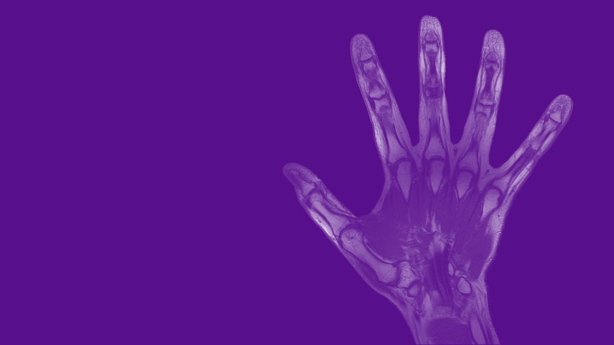
CAI2R creates technologies for better acquisition, reconstruction, and analysis of medical images.
Our innovations advance research in biomedicine and our best technologies become leading-edge tools in clinical radiology.
October 21-22, 2026
Join us at NYU Langone Health for the 2026 i2i Workshop dedicated to learning: from learning mechanisms in humans to machine learning strategies in image acquisition, reconstruction, analysis, and AI-oriented hardware.
CAI2R (pronounced care) is a National Center for Biomedical Imaging and Bioengineering supported by the National Institute of Biomedical Imaging and Bioengineering (NIBIB) and operated by NYU Langone Health.
A Unique Model for Academic Medical Research
Research and development in biomedical imaging are extraordinarily complex. We assemble translational research teams that meet the challenge.
Technological Innovation
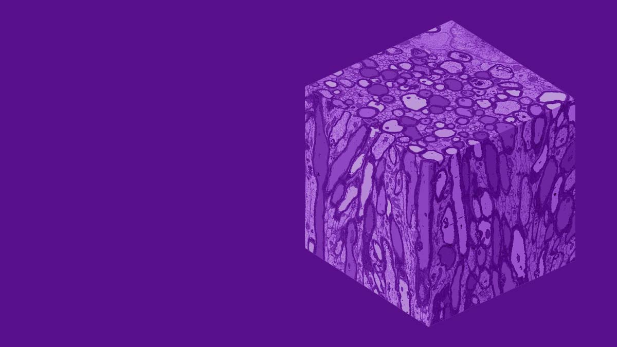
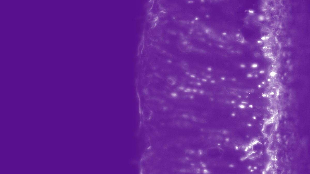
CAI2R research leads the field in fast imaging, machine learning for image acquisition and reconstruction, ultraflexible biomorphic hardware, complementing MRI data with novel sensing strategies, mapping of tissue microstructure, and artificial intelligence methods for early detection of disease.
Interdisciplinary Collaboration
Our Center brings together basic scientists, engineers, clinical radiologists, physicians, computer scientists, and specialists from the medical imaging industry. We form innovative research partnerships with institutions in medicine, academia, and tech.
Clinical Translation
CAI2R innovations advance knowledge, diagnosis, and therapy of neurological conditions, musculoskeletal conditions, cardiovascular conditions, and cancer. Our research is focused on scientific and clinical applications of new biomedical imaging technologies.
Open Science
CAI2R shares research software and data resources in order to encourage progress throughout the field. Our training activities include hosting visiting scientists, a regular radiology research forum, and a biennial workshop devoted to emergent imaging technologies.

Latest News
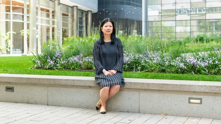
Linda Moy, radiologist and researcher whose work spans breast imaging and artificial intelligence, talks about patient-centric radiology, AI systems in clinical practice, and the field's rising environmental awareness.
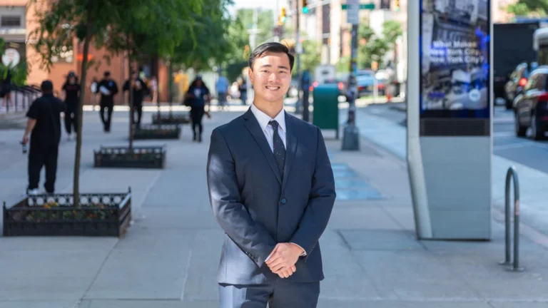
Congratulations to Peter Hsu on a successful defense of his doctoral dissertation in biomedical imaging and technology at NYU Grossman School of Medicine.
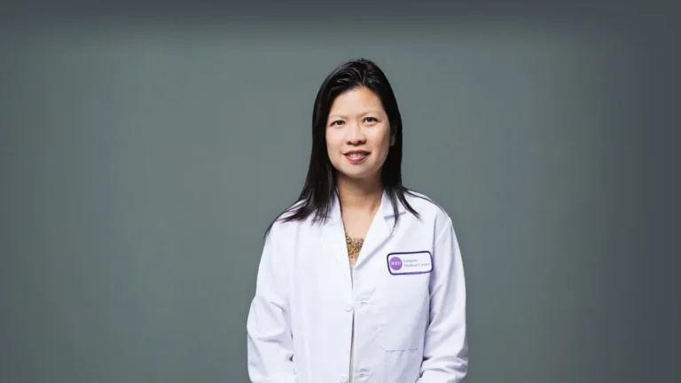
Congratulations to Linda Moy on becoming the 2026–2027 vice president–elect of the International Society for Magnetic Resonance in Medicine.
Latest Resources
Denoising multi-echo spin-echo MRI data with Marchenko-Pastur principal component analysis.
Quantitative mapping of myelin content using multi-T2-component fitting of multi-echo spin-echo MRI data.
Software for quantification of T2 relaxation time and proton density from multi-echo spin-echo MRI data.
Software for estimation of multiple variations of the cumulant expansion from diffusion MRI data.

A neural network for reconstructing electrical properties of materials and biological tissue from MRI data, plus synthetic phantoms.
By the Numbers
scientific publications
resource downloads
workshop participants
team members
collaborative projects
services projects
patents
years since founding