
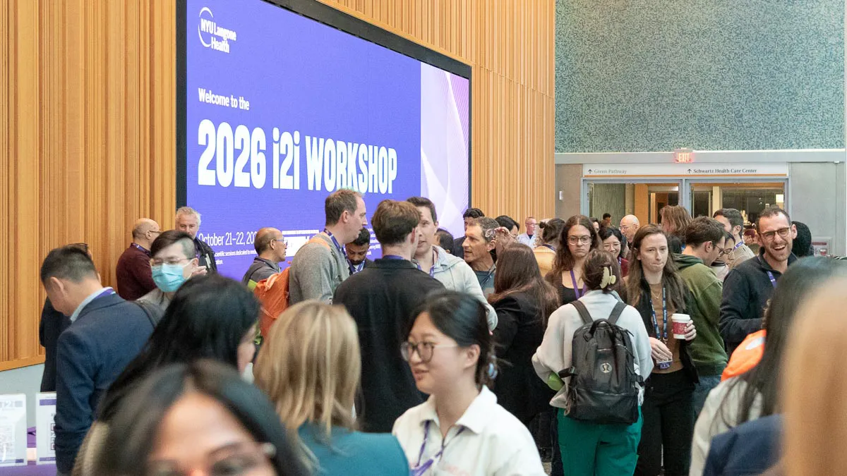
On October 21 and 22, more than 220 experts in radiology, imaging research, machine learning, and neuroscience convened at NYU Langone Health for the fourth edition of the i2i Workshop. The conference, named after the tagline “from innovation to implementation in imaging,” is held every few years by the Center for Advanced Imaging Innovation and Research to explore the emerging frontiers in biomedical imaging science.
“We wanted to bring together people from different disciplines and fields to understand what learning means in biology, what it means in machine learning and in biomedical imaging,” said Riccardo Lattanzi, PhD, professor of radiology and director of the Bernard and Irene Schwartz Center for Biomedical Imaging at NYU Langone, who co-chaired the workshop.
“The meeting theme is well aligned with our institutional and departmental vision of being a learning healthcare system,” said Hersh Chandarana, MD, professor of radiology and urology at NYU Grossman School of Medicine and vice chair for innovation and predictive diagnostics in the department of radiology at NYU Langone.
Dr. Chandarana, who co-chaired the meeting and is the overall principal investigator at the Center for Advanced Imaging Innovation and Research, explained that in a large health system, a lot of the data created in the course of providing care remain underutilized. The question, he said, is how to systematically turn these data into knowledge to better guide care and improve patient outcomes.
A Decade of i2i
Since its launch in 2016, the i2i Workshop series has reflected—if not anticipated—the evolution of machine learning methods in biomedical imaging research. The inaugural sessions featured talks on deep learning–based image reconstruction, a then cutting-edge innovation that NYU Langone scientists were among the first to propose.
The field’s intense interest in machine learning was captured in the 2018 i2i Workshop, with sessions such as “Reimagining MR Scanners,” “Rethinking RF Coils,” and “The AI Revolution.”
The 2023 meeting reflected a phase in which AI had begun to enable advances across the imaging domain: from ultra-high to ultra-low field MRI; from data acquisition to image formation to image analysis; across multiple imaging modalities; even reaching toward new, supplemental sensing technologies.
The 2026 workshop highlighted a yet different stage, with AI becoming increasingly woven into the fabric of imaging research, radiology practice, and patient care. Hence, the invitation to reflect on machine learning and learning itself.
Learning in Biomedical Imaging
Across both workshop days, the scientific sessions illuminated AI applications in imaging research and asked what we stand to learn from them.
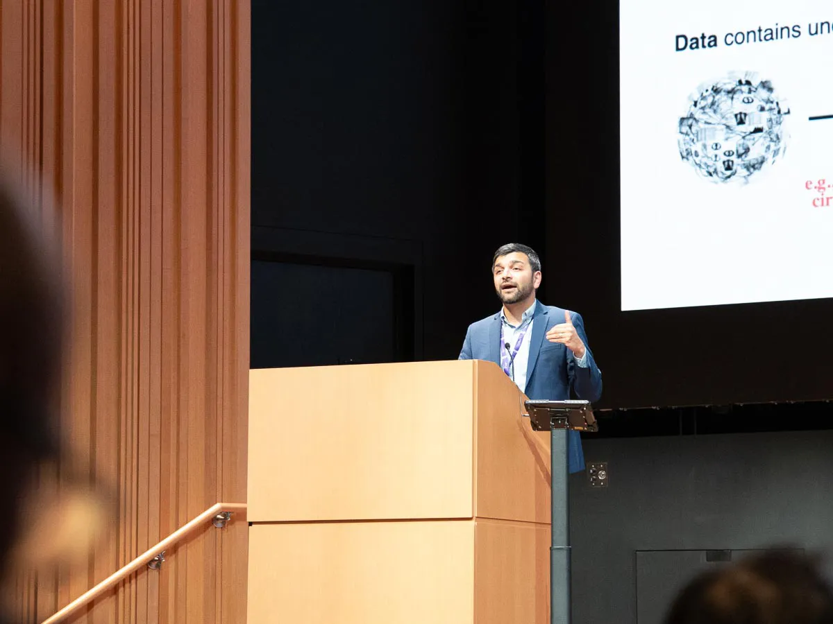
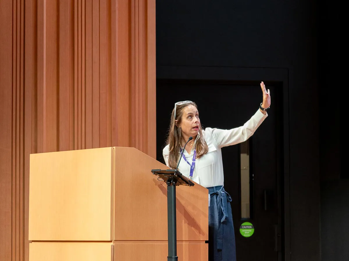
“For me, learning is an iteration process,” said Fuyixue Wang, PhD, assistant professor at Harvard Medical School and Massachusetts General Hospital, who spoke about advances in brain imaging that bring new information to both medical applications and neuroscience. “The clinical or neuroscientific problem drives the innovation in terms of developing new technology, and then new technologies create new data sets that inspire new questions,” said Dr. Wang in an interview. Those questions, in turn, “inspire the next generation of imaging technologies,” she said.
Akshay Chaudhari, PhD, associate professor of radiology and biomedical data science at Stanford University, discussed the promise of multimodal foundation models capable of learning from a wide range of data types: clinical notes, radiology reports, medical images, medical textbooks, scientific literature, demographic data, genetic information, and data from wearables. Foundation models have shown potential to generalize well from training datasets to new cases. In an interview, Dr. Chaudhari raised the possibility that AI models trained on multimodal medical data could both improve care and guide discovery. “Instead of just being able to predict future disease activity or be able to diagnose specific findings, perhaps the existence of this data at this scale can allow us to learn something new about the underlying biology itself,” he said.
At NYU Langone, researchers are investigating whether AI screening of medical images for conditions beyond the original reason for an exam can benefit patients. Miriam Bredella, MD, MBA, the Bernard and Irene Schwartz Professor of Radiology at NYU Grossman School of Medicine, vice chair for strategy in the Department of Radiology at NYU Langone Health, talked about a system studied at NYU Langone that flags suspected osteoporosis on abdominal and chest CT scans taken for unrelated reasons—an approach called opportunistic imaging.
“Actually, opportunistic imaging is not new at all,” said Dr. Bredella, who is also the associate dean for translational science at NYU Langone and director of the NYU Clinical and Translational Science Institute. However, seizing these opportunities has until recently required expert labor. “It wasn’t really possible to do this real-time or at large scale,” Dr. Bredella said. “Now, having AI, we can do all of this within seconds and automatically.” Osteoporosis screening is just one example of how mining existing images can yield early health warnings. Similar flags are conceivable for signs of cardiovascular disease, metabolic disease, and other conditions. “And that brings radiology more toward prevention,” said Dr. Bredella.
Contemplating How Humans Learn
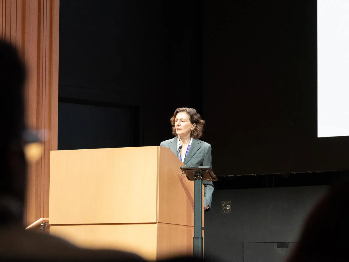
Little of what we know about learning in living organisms directly corresponds to what is meant by learning in AI, said Cristina Alberini, PhD, the Julius Silver, Roslyn S. Silver and Enid Silver Winslow Professor with the Center for Neural Science at New York University, in her talk on the biology of learning and memory. In an interview, Dr. Alberini said, “I don’t see the correspondence to artificial learning, machine learning, storage of information that is used in those systems,” even though both domains use similar vocabulary. For that reason, she said, “It is extremely important to be in the same room, possibly because novel ideas will come out from there in both directions, but especially in the direction of creating systems that learn better.”
In a talk devoted to what happens when we experience difficulty learning, Nadine Gaab, PhD, the Silvana Chris Pascucci Professor in Learning Differences at Harvard University, shared insights from longitudinal studies that seek to deepen the knowledge of how children learn to read, write, and do math. In an interview, Dr. Gaab, whose research incorporates magnetic resonance imaging and aims at informing educational policy, said that “Usually, we only think of the translational aspect as being from the bench to the bedside, but we have created a lot of impact from the MRI bed to the school bench,” showing that technical advances in MRI and image analysis “can impact audiences beyond the hospital.”
Dr. Gaab drew a parallel to preventive medicine with the idea that a child’s risk of developing a learning disability could be identified early in the course of regular pediatric care, and addressed in a way that reduces the likelihood of having to seek a special educator’s intervention later. “We call this preventative education, and we think it will have tremendous impact on academic outcomes, mental health outcomes, but also economic and vocational outcomes,” she said.
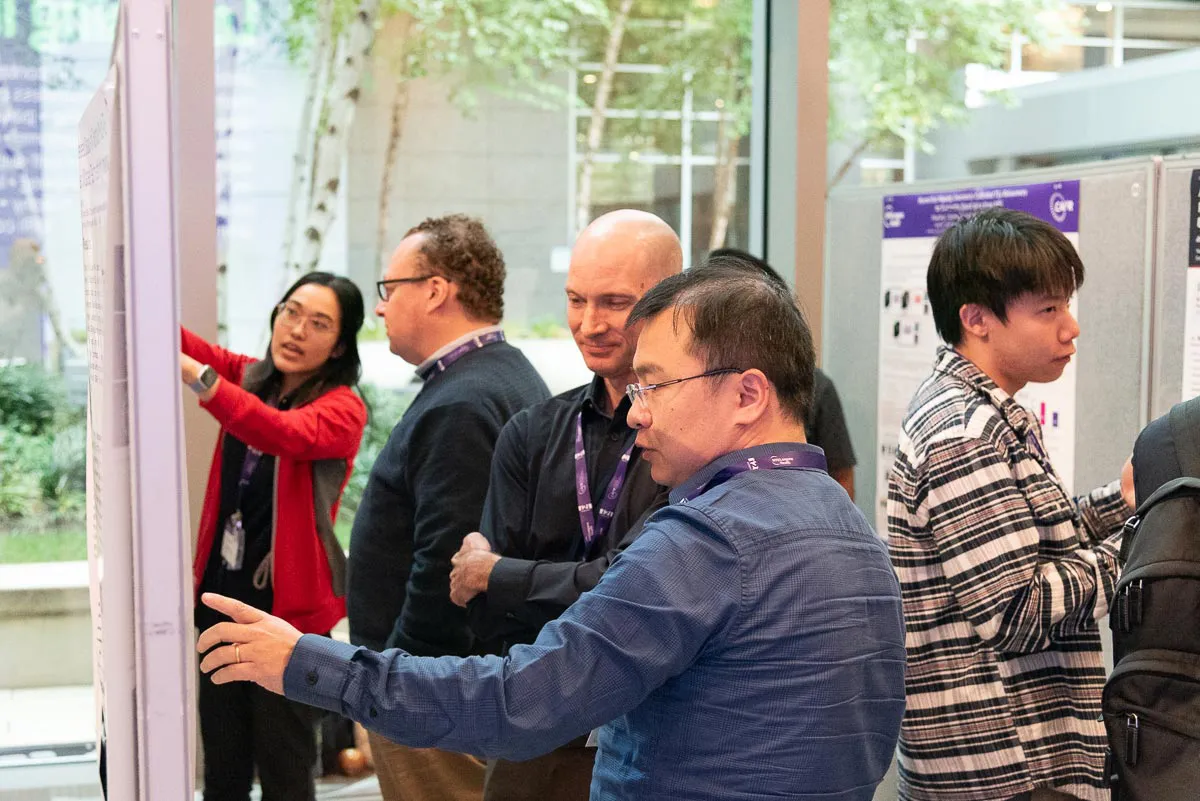
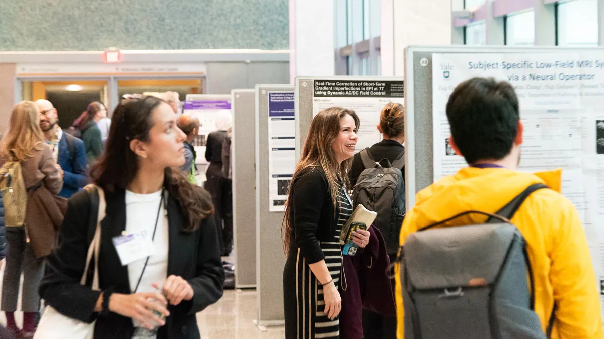
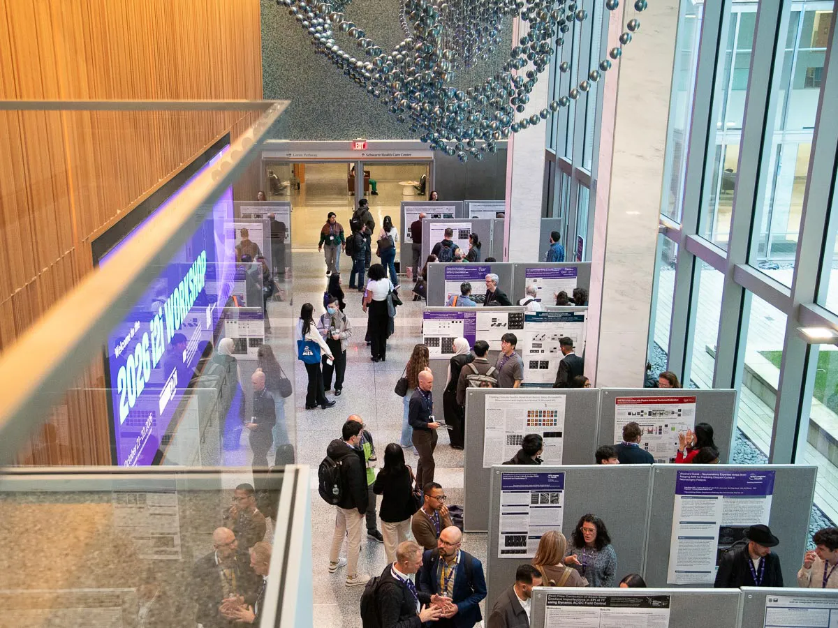
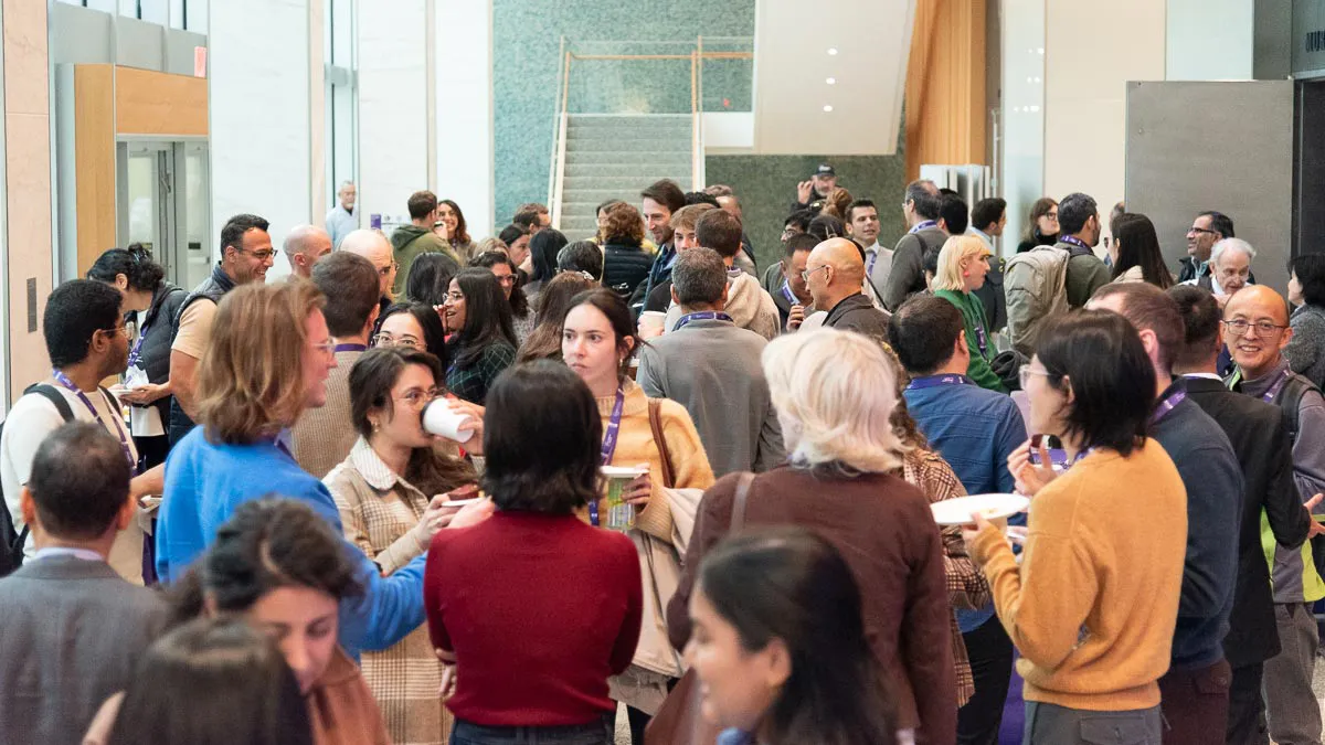
The workshop also included a poster session, evening receptions, and breaks that gave participants time to connect and exchange ideas.
“The bet behind the workshop is simple,” said Dr. Lattanzi. “That placing the mechanism of human learning beside the mechanism of machine learning in a room full of imaging scientists generates new questions.”
Related Stories

How a moonshot effort to build an open-source MRI scanner from scratch in less than a week played out, and what it means for imaging research at large.

In a return from a pandemic hiatus, the i2i Workshop encouraged a broad and varied look at what imaging is and where it's headed.