

In a new study published in the journal Investigative Radiology, researchers with the Department of Radiology at NYU Langone Health, the Bernard and Irene Schwartz Center for Biomedical Imaging, and the Center for Advanced Imaging Innovation and Research measure the effect of high acceleration rates on the quality of brain MRIs reconstructed with deep learning algorithms. The authors have found that at accelerations sixfold and greater MRI regions that contain abnormal features deteriorate more than do image regions that appear normal, a divergence that widens as the magnitude of the speedup grows.
“People know that this can occur, but it has been somewhat anecdotal,” said Yvonne Lui, MD, professor and vice chair for research in radiology at NYU Langone. “This paper rigorously quantifies that.”
Using nearly 5,000 brain MRI exams from NYU Langone’s open-source fastMRI dataset, the scientists trained and validated a deep learning image reconstruction network at retrospective acceleration factors of 2, 4, 6, 8, 10, and 12. Twofold acceleration refers to generating an image from half the amount of raw data that would be acquired for a fully sampled MRI; fourfold acceleration means generating an image from a quarter of the underlying data; and so on. The research team then tested the models on a separate set of 1,000 brain exams complemented by radiologist annotations obtained from an open-source project called FastMRI+.
The annotations made it possible for the researchers not only to benchmark whole accelerated MRIs against fully sampled references, but also to compare regions of salient clinical interest—local abnormalities and pathologies—to similar but normal-appearing regions in other images from the test set.
To gauge image quality, the authors employed three metrics of the similarity of a given MR image to its reference: normalized mean square error (NMSE), peak signal-to-noise ratio (PSNR), and structural similarity index measure (SSIM). An analysis of these measures numerically confirms “disproportionate loss of image quality in areas of abnormality at high acceleration factors,” write the authors, referring to speedups sixfold and greater.
“Prior studies evaluate globally,” said the lead author Shengjia Chen, MS, now a doctoral candidate in computational pathology at the Icahn School of Medicine at Mount Sinai, who got involved in the investigation while studying biomedical informatics at NYU Grossman School of Medicine.
But whole-image analysis leaves open the question of “whether image quality degrades evenly as acceleration increases,” he said. In the new study, “we separate each slice into abnormal regions and normal regions, and do a quantitative study and a statistical analysis to show the differences.”
Patricia Johnson, PhD, assistant professor of radiology at NYU Grossman School of Medicine and the corresponding author on the study, said part of the motivation was “to encourage caution for using deep learning reconstruction with very high acceleration factors.”
Acceleration and Patterns
In contemporary MRI, making scans faster means finding clever ways of forming images from fewer measurements, and artificial intelligence algorithms trained on large numbers of MRIs have proven effective at accurately generating complete images from incomplete underlying data. But this ability tends to break down at high acceleration rates—when raw data become scant.
One striking characteristic of deep learning–based reconstruction is that such breakdowns are not obvious in the resulting images. There are no shadows, auras, ghosting, banding, streaking, or other visual cues known to radiologists as artifacts of imperfect image formation. As a result, “model outputs are presented with extreme certainty even in areas of clearly uncertain image rendering,” write the authors of the study.
“It’s called pseudo-normalization,” said Chen. “When we use higher acceleration rates, we see normalization issues: the model pretends to reconstruct the regions as normal.”
Pseudo-normalization is not itself new—in a 2022 study that explored the limits of AI-based reconstruction by pushing acceleration factors as high as a hundredfold, researchers from NYU Langone, Facebook AI Research, Stealth, and Friedrich-Alexander University Erlangen-Nürnberg defined this phenomenon as “an effect of the model that makes an abnormal brain appear normal.” Dr. Lui said it can be thought of as the opposite of hallucination.
“Abnormal scans and abnormal areas—tumors, areas of concern—are actually more difficult for deep learning reconstructions to render than normal scans,” said Dr. Lui, who is also a practicing neuroradiologist. “Of course that’s exactly what we need to see: abnormal areas.”
Why does pseudo-normalization happen?
Dr. Johnson outlined the mechanism behind this behavior: the less raw data there is to work from, the more the model tends to fall back on its training, and because abnormal regions tend to be less represented in training data, a deep learning model is less likely to accurately predict abnormalities in a heavily undersampled scan. “The takeaway is that when you push acceleration very high, the model leans on that prior more heavily and you start to disproportionately lose information from abnormal regions,” she said.
Trust over Speed
The findings do not question the potential of deep learning–based image reconstruction as a method, but they do indicate that more research is needed to evaluate new models.
“It’s important to understand the boundaries,” said Dr. Lui, emphasizing that the investigation explored a range of speedups significantly greater than the twofold and fourfold accelerations currently approved for select applications in clinical radiology. “In this study, we really do push the boundaries—it’s extremely high levels of acceleration.”
Dr. Johnson said that quantification is just the first step.
“These metrics are commonly used and they’re great for development,” Dr. Johnson said of the NMSE, PSNR, and SSIM measures used in the study. “They’re great as a starting point, but they don’t necessarily capture so well when you’re losing very small features that are clinically relevant,” she said. “It’s really only one of the steps in the evaluation pipeline.”
The authors call for qualitative human-reader studies to provide more information about the clinical significance of the deterioration of representations of local abnormalities. They also note the need for prospective studies evaluating new, accelerated deep learning–based reconstruction models.
One early example is a 2020 study by imaging researchers at NYU Langone and Facebook AI Research that demonstrated diagnostic interchangeability of MRIs reconstructed with deep learning at fourfold acceleration with conventionally reconstructed counterparts.
It’s interchangeability and non-inferiority studies of this kind that built trust and enabled clinical adoption of twofold and fourfold accelerated deep learning reconstructions. Dr. Johnson said that as the technology advances, such assessments should be done for every model, acceleration, and application area.
“We’ve done the studies and we know fourfold acceleration for knee [MRI] is reliable, but we don’t know whether tenfold for knee is going to give you the same diagnostic information,” she said. “Evaluation needs to be acceleration factor–dependent and anatomy-specific.”
Understanding the limitations, “leads us to try to do better,” said Dr. Lui, “to try to identify areas where the reconstruction is not as good, and how we can improve the technology to ensure better data fidelity and to render the image more justly.”
This story was published on September 30, 2026, and augmented the following day.
Research reported in this story is supported by the National Institute of Biomedical Imaging and Bioengineering under award number R01EB024532 and P41EB017183. The content is solely the responsibility of the authors and does not necessarily represent the official views of the National Institutes of Health.
Related Resource
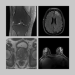
Raw k-space data and DICOM images from thousands of MRI scans of the knee, brain, and prostate, curated for machine learning research on image reconstruction.
Related Stories

The fastMRI dataset now includes curated breast MRI data to boost AI innovation in radial, dynamic contrast-enhanced, ultrafast MRI of the breast.

Radhika Tibrewala, graduate student in biomedical imaging, talks about the new fastMRI prostate dataset, deep learning in MRI, and how she started a PhD remotely in 2020.

Patricia Johnson, who researches machine learning image reconstruction, talks about faster MRI, visual preferences, and diagnostic interchangeability.