
Imaging scientists at NYU Langone Health are proposing to improve prostate MRI by removing anatomical distortion, increasing image resolution, and shortening scan time of a key component of exams commonly used to evaluate the likelihood and severity of cancer. The National Institute of Biomedical Imaging and Bioengineering (NIBIB) is supporting the project with up to $1 million over five years awarded in June to Jingjia Chen, PhD, postdoctoral fellow at NYU Grossman School of Medicine and researcher with NYU Langone’s Center for Advanced Imaging Innovation and Research. The award is a K99/R00 grant, intended to “facilitate a timely transition of outstanding postdoctoral researchers … to independent, tenure-track or equivalent faculty positions,” according to the NIBIB.
For most patients, the path to prostate cancer diagnosis begins with a blood assay for prostate-specific antigen (PSA) and ends with a biopsy, a procedure in which tissue samples are extracted with a needle probe and sent to a laboratory for analysis. But only about one in every five men with high PSA levels is ultimately found to have clinically significant prostate cancer. Hence, doctors rely on a variety of additional tests to narrow down which patients are at high risk of malignancy before recommending that tissue be sampled. In this middle ground between the frontline and the final tests, MRI is increasingly helping guide decisions on whom to send home and whom to biopsy. Imaging has a specificity rate of approximately 50 percent—not perfect but meaningfully higher than that of a PSA assay—and in 2026 radiologists in the U.S. will have read hundreds of thousands of prostate MRIs.
An MRI of the prostate can comprise several types of scans, but the most useful diagnostic information comes from two that are always included: T2 “structural” imaging and diffusion-weighted imaging (DWI). The T2 images have high resolution and offer fine anatomical detail; the DWI—so named for its ability to probe the molecular motion of water inside the body—is coarser but shows areas where diffusion is restricted, signaling possible malignancy. It’s this diffusion component that the project led by Dr. Chen centers on.
“DWI is very important in the prostate cancer diagnostic pipeline,” said Dr. Chen. Other imaging sequences, such as a coronal structural scan or a dynamic contrast-enhanced scan, can provide additional nuance but aren’t essential for every patient, “but you cannot miss DWI.”
Hersh Chandarana, MD, professor of radiology and urology at NYU Grossman School of Medicine and overall principal investigator at the Center for Advanced Imaging Innovation and Research at NYU Langone, explained that “you can think of diffusion as providing specificity.” For example, “you may see some abnormality on T2, and the question is: is this abnormality cancer or not? We rely on diffusion imaging to help make that decision.” He added that in the area of the prostate called the peripheral zone, diffusion images lead and T2 images support the evaluation. This T2-plus-DWI combination is the backbone of the standard prostate MRI, also called biparametric.
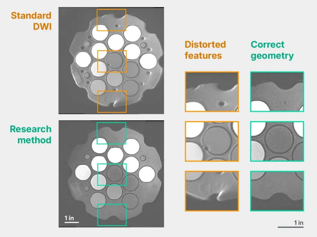
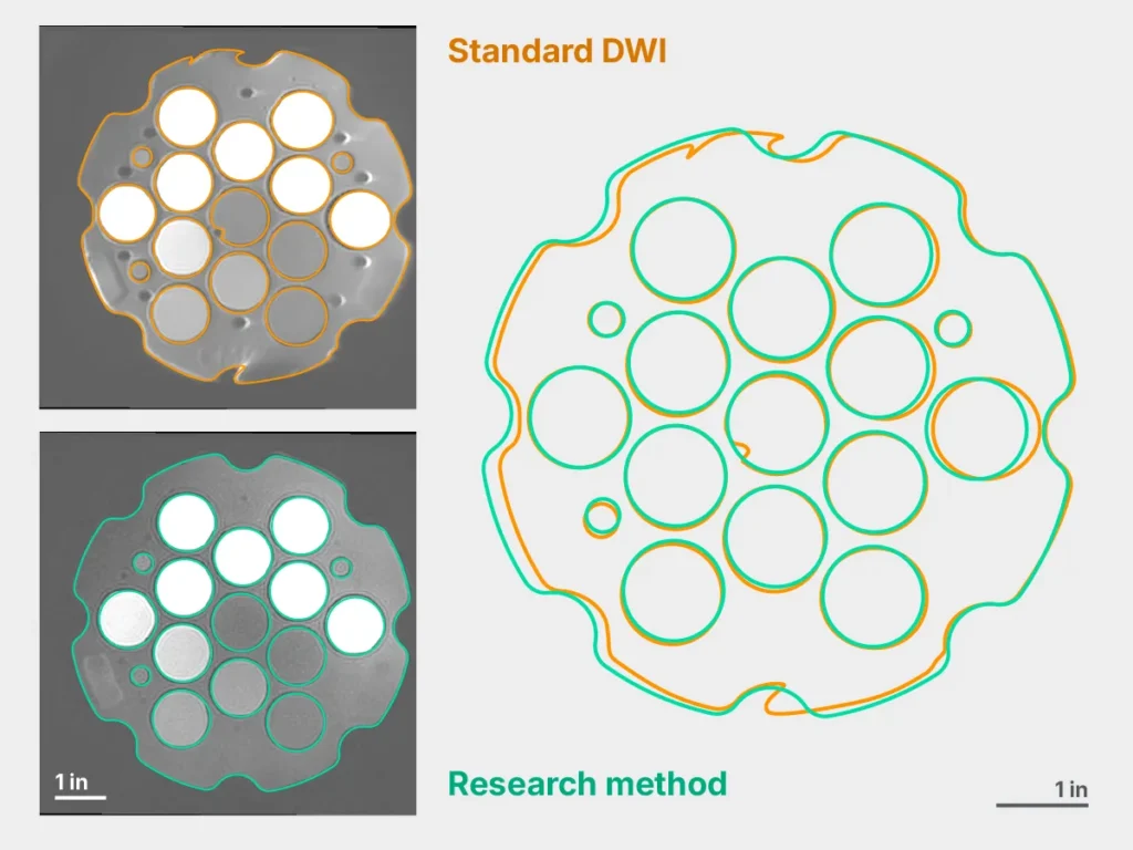
But the standard diffusion-weighted images have relatively low resolution and are often marred by distortions that appear as kinks and warps in the anatomy. The deformities show up the most in the peripheral zone of the prostate, exactly where DWI guides diagnosis.
Radiologists get around these flaws by cross-referencing the diffusion-weighted images with the T2 structural scans, which do not deform, and comparing anatomical landmarks. “It is working but it’s suboptimal,” said Dr. Chen.
Dr. Chen is proposing to develop anatomically accurate, high-resolution, fast DWI—a combination that, if successful, has the potential to reshape the biparametric prostate MRI exam. Her project is mentored by Dr. Chandarana; Li Feng, PhD, associate professor of radiology at NYU Grossman School of Medicine; and Daniel Sodickson, MD, PhD, chief medical scientist at Function Health and adjunct professor of radiology at NYU Grossman School of Medicine.
A Small Organ in a Crowded Neighborhood
MRI does best with large, fleshy, relatively uniform structures that don’t move around much. The brain or the thigh muscle, for example, make excellent scan targets. The prostate does not. Located in crowded terrain, with the bladder sitting atop, the colon pressing from behind, and the urethra tunneling through, the prostate is surrounded by pockets of urine and gas, which have disparate magnetic properties. The juxtaposition of areas rich in MRI signal with those starved of it leads to magnetic susceptibility artifacts—distortions. (In patients who have metallic hip implants, the magnetic environment becomes even more complex.)
These artifacts, in turn, limit the resolution of DWI because scanning longer to acquire more detail tends to exacerbate the flaws. It doesn’t help that the organ is small, about the size of a walnut—though it can become enlarged if not healthy—and cancerous lesions inside it can be tiny.
Compounding the challenge, the prostate and its immediate surroundings experience a fair bit of motion: not only due to the flows of the nearby liquids and gases but also because of the pulling and pushing of the diaphragm, which affects soft tissues in the abdomen all the way down to the pelvic floor.
“There’s a lot of stuff going on in the pelvic region,” said Dr. Chen. The difficult conditions present an opportunity for a better imaging method to make a substantial difference in the clinic.
Known Ingredients, Novel Recipe
In order to unkink prostate DWI, the researchers are proposing to change the way the diffusion-weighted signal is received by combining two methods known as spin echo and gradient echo. Dr. Chen explained that “spin echo is robust against magnetic susceptibility effects”—free from distortion—“but slow and blurry.” To counteract the downsides, “we add some gradient echo,” a technique similar to that used in the current clinical standard, prone to flaws but fast. By interleaving these readout methods, the research team aims at balancing quality with speed.
The proposed approach also incorporates a type of radial sampling known as PROPELLER. One of the characteristics of radially sampled data is that every acquired “blade” goes through the central region of k-space, MRI’s raw-data dimension. The meeting of lines in the center creates a shared reference that can be used as a basis for motion correction during image reconstruction.
To increase resolution and shorten scans, the team plans to train deep learning algorithms to reconstruct images from raw DWI data. The researchers also plan to study whether the method improves diagnosis and whether quantitative metrics derived from DWI correlate with biopsy analyses.
Although the respective elements of the proposed imaging approach are not themselves new, the envisioned advance resides in combining them all to solve an unmet need in a diagnostic tool of growing import. “It’s a whole design of acquisition and reconstruction,” said Dr. Chen.
Early research led by Dr. Chen on this method earned two recognitions in 2025: one at an International Society for Magnetic Resonance in Medicine workshop dedicated to body MRI, another at an international symposium devoted to magnetic resonance in radiation therapy.
Dr. Chandarana said that distortion-free, high-resolution diffusion-weighted imaging of the prostate “would have multiple important contributions” with potential knock-on effects on the whole exam.
DWI of the prostate is acquired at two diffusion weightings called b-values—“a low b-value, which is a T2-like contrast, and a high b-value, which is a diffusion contrast,” explained Dr. Chandarana. “If we had robust imaging at low and high b-value, I might ask myself if I need T2 imaging.” In this hypothetical scenario where diffusion-weighted imaging suffices, the biparametric MRI exam reduces to a DWI scan. It’s conceivable that an appointment that currently takes about a half hour could drop to as little as 10 minutes, lowering costs and broadening availability. “That’s the end game,” said Dr. Chandarana.
Though the aims of the research project led by Dr. Chen do not extend that far, she cites the potential clinical impact as a strong motivation. “I come from a technical background and am still training in a technical field,” said Dr. Chen. “I want to make sure that I’m learning to think in a way that helps me build something useful.”
Research reported in this story is supported by the National Institute of Biomedical Imaging and Bioengineering under award number K99EB039095. The content is solely the responsibility of the authors and does not necessarily represent the official views of the National Institutes of Health.
Related Stories

Jingjia Chen, postdoctoral fellow who investigates longitudinal imaging and imaging in the presence of motion, talks about using the past in the present, improving body MRI, and taking better pictures.

Congratulations to Jingjia Chen and coauthors on winning second prize in oral presentations at the International Society for Magnetic Resonance in Medicine workshop on body MRI.