Hersh Chandarana, MD, and Yulin Ge, MD, imaging scientists at NYU Langone Health, have been named senior fellows of the International Society for Magnetic Resonance in Medicine (ISMRM). The honor celebrates “those who have made significant and substantial contribution to research in a field within the Society’s purposes, … to the development of the Society, … [or] to education in [magnetic resonance].” Drs. Chandarana and Ge are among 17 researchers distinguished with senior fellowship in 2025 announced during the annual meeting of the ISMRM held May 10-15 in Honolulu.
Dr. Chandarana—professor in the departments of radiology and urology at NYU Grossman School of Medicine; vice-chair for radiology hospital operations and analytics at NYU Langone Health; and co-director for clinical and translational research at NYU Langone’s Center for Advanced Imaging Innovation and Research—has been cited by the ISMRM for “making abdominopelvic MR imaging more accessible, motion robust, free-breathing, fast and easy by developing novel free-breathing and accelerated MRI methods for motion robust and motion-sorted imaging.”
“I’m truly grateful for this recognition and for the community that has supported and shaped my journey,” said Dr. Chandarana, adding that the ISMRM “has felt like a professional home to me said the society continues to be “a source of inspiration, growth, and connection.”
Dr. Chandarana is one of the creators of GRASP, an MRI method that delivers sharp images without requiring patients to perform repeated breath holds in the scanner. GRASP MRI, which combines radial sampling, parallel imaging, and compressed sensing techniques, was invented about a decade ago at the Center for Advanced Imaging Innovation and Research by Ricardo Otazo, PhD; Li Feng, PhD; Tobias Block, PhD; Leon Axel, MD, PhD; Daniel Sodickson, MD, PhD; and Dr. Chandarana. Since then, the method has been used in more than 150,000 clinical exams at NYU Langone and reached radiology clinics around the world as a product shipped with Siemens MRI scanners.
GRASP has also found applications beyond abdominal and pelvic imaging, including in cardiac imaging, neuroimaging, and breast imaging, and has become foundational to a growing compendium of extensions that incorporate the latest advances in image acceleration and machine learning. The method and its continued development exemplify how the Center for Advanced Imaging Innovation and Research is leading the field toward faster, more continuous, more comprehensive, and more intelligent MRI.
Dr. Ge, professor in the department of radiology at NYU Grossman School of Medicine, has been recognized by the ISMRM “for pioneering contributions to the development of biomarkers in aging and neurodegenerative disorders using anatomic, physiological, and functional MRI techniques.”
“I am deeply honored to receive this recognition from … the world’s largest community driving innovation, clinical translation, and education in MRI,” said Dr. Ge, calling the award a “milestone that reflects the incredible dedication of my team and collaborators” and a “deep commitment to pushing the boundaries of advanced MRI to better understand neurodegenerative diseases.”
Major contributions include the discovery of what is now known as “central vein sign,” a distinct pattern of lesions in the brain that is associated with multiple sclerosis, first observed by Dr. Ge and colleagues at NYU Langone’s Center for Biomedical Imaging in 2008; and the development that same year of TRUST, an MRI technique for measuring the brain’s oxygen metabolism, co-authored by Dr. Ge and Hanzhang Lu, PhD. Both advances have since proven valuable in diagnosing and studying multiple sclerosis.
More recently, Dr. Ge’s research has focused on investigating vascular contributions to cognitive impairment and dementia, an area of inquiry known as VCID. Together with colleagues at NYU Langone and elsewhere, Dr. Ge has developed MRI methods that enable noninvasive in vivo study of hair-thin arteries in the brain and is exploring age-related tortuosity—or curvature—of blood vessels. The work has shown that arteries of all sizes tend to become more tortuous with age, a change that may result in less efficient delivery of oxygen. In 2022, Dr. Ge served as president of the International Society for Neurovascular Disease, and in 2024 he was inducted into the college of fellows of the American Institute for Medical and Biological Engineering. A profile published on this blog in 2024 describes his scientific path from a fascination with early MRI machines to discoveries made with ultra-high-field scanners.
Related Stories

The fastMRI dataset now includes curated breast MRI data to boost AI innovation in radial, dynamic contrast-enhanced, ultrafast MRI of the breast.

Yulin Ge, imaging researcher at NYU Langone, originally trained to be a radiologist. A fascination with MRI has led him to pursue science that illuminates aspects of neurological health, disease, and aging.

Congratulations to Els Fieremans and Dmitry Novikov on being named among the 2024 senior fellows of the International Society for Magnetic Resonance in Medicine.
Related Resources

Respiratory motion compensation, automatic bolus timing, and coil-weighted unstreaking for GRASP.
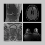
Raw k-space data and DICOM images from thousands of MRI scans of the knee, brain, and prostate, curated for machine learning research on image reconstruction.
-
A reconstruction method that sorts dynamic data into motion states in accelerated MRI.
-
Reconstruction code for fast and flexible free-breathing dynamic volumetric MRI.