We are sharing code for respiratory-weighted, aortic contrast enhancement-guided, and coil-unstreaking (RACER) features integrated with golden-angle radial sparse parallel (GRASP) reconstruction of MR images.
RACER extends GRASP with new functionality: respiratory motion compensation, automatic bolus timing, and coil-weighted unstreaking for imaging of the liver.
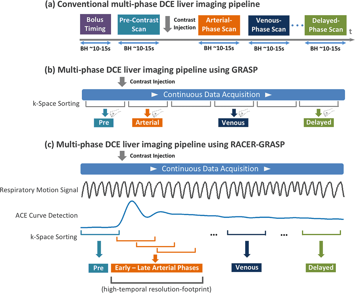
The illustration below shows a comparison of conventional, GRASP, and RACER-GRASP workflows for dynamic contrast enhanced liver imaging.

(a) In conventional multiphase DCE liver imaging, separate 3D image volumes are acquired in desired contrast-enhancement phases during multiple breath-holds. A bolus timing step is performed before the DCE scan to ensure optimal detection of desired contrast phases.
(b) In multiphase DCE liver imaging using GRASP, data are continuously acquired under free breathing, and images can be reconstructed with flexible temporal resolutions by grouping a specific number of consecutive spokes as each contrast phase. However, the optimal detection of desired contrast-enhanced phases is not guaranteed, and radiologists must manually select their desired contrast phases.
(c) In multiphase DCE liver imaging using RACER-GRASP, a respiratory motion signal and an aortic contrast enhancement (ACE) signal are extracted from the acquired k-space to guide k-space sorting and image reconstruction. ACE information can ensure optimal detection of desired contrast phases.
For more details, see the related publication below.
Get the Code
The software available on this page is provided free of charge and comes without any warranty. CAI²R and NYU Grossman School of Medicine do not take any liability for problems or damage of any kind resulting from the use of the files provided. Operation of the software is solely at the user’s own risk. The software developments provided are not medical products and must not be used for making diagnostic decisions.
The software is provided for non-commercial, academic use only. Usage or distribution of the software for commercial purpose is prohibited. All rights belong to the author (Li Feng) and NYU Grossman School of Medicine. If you use the software for academic work, please give credit to the author in publications and cite the related publications.
Contact
Questions about this resource may be directed to Li Feng, PhD, at Li.Feng@nyulangone.org.
Related Resources
A reconstruction method that sorts dynamic data into motion states in accelerated MRI.
Reconstruction code for fast and flexible free-breathing dynamic volumetric MRI.
Related Post
Li Feng, developer of fast MRI techniques, talks about going beyond speed, his path to academia, and the rewards of persistence.


