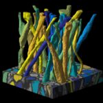
We are making available segmentation code and scanning electron microscopy (SEM) data used to characterize inner axon diameters and fiber orientation dispersion in a sample of murine corpus callosum.
We have used these tools and data to examine assumptions that underlie the practice of modeling axons as perfect cylinders in diffusion MRI (dMRI). Our findings indicate that such modeling may be too simplistic for dMRI in the brain. For more detail, see the related publication.
The SEM data include:
- datac.nii (a stack of SEM data, 200 slices; resolution: 24nm by 24nm by 100nm; volume: 36μm by 48μm by 20μm)
- fibers.nii (intra-axonal space segmented by using random-walker-based approach)
- maskc.nii (foreground mask)
- myelin_mask.nii (myelin mask generated by pixel-wise classifier in ilastik)
The code for random walker (RaW) segmentation and quantification of fiber orientation and axonal diameter is maintained on Github.
Related Publication
Along-axon diameter variation and axonal orientation dispersion revealed with 3D electron microscopy: implications for quantifying brain white matter microstructure with histology and diffusion MRI.
Brain Struct Funct. 2019 May;224(4):1469-1488. doi: 10.1007/s00429-019-01844-6
Please cite this work if you are using the intra-axonal space segmentation from 3D SEM of murine corpus callosum in your research.
Get the SEM Data
The software available on this page is provided free of charge and comes without any warranty. CAI²R and NYU Grossman School of Medicine do not take any liability for problems or damage of any kind resulting from the use of the files provided. Operation of the software is solely at the user’s own risk. The software developments provided are not medical products and must not be used for making diagnostic decisions.
The software is provided for non-commercial, academic use only. Usage or distribution of the software for commercial purpose is prohibited. All rights belong to the authors (Hong-Hsi Lee) and NYU Grossman School of Medicine. If you use the software for academic work, please give credit to the author in publications and cite the related publications.
Get the RaW Code
To complement the SEM data, we are sharing code for random walker (RaW) segmentation and quantification of fiber orientation and axonal diameter.
Contact
Questions about this resource may be directed to Hong-Hsi Lee, PhD at hlee84@mgh.harvard.edu.
Related Post
Scientists at NYU Langone Health show that MRI signal can detect axonal features long assumed to lie beyond the reach of magnetic resonance imaging.