Radhika Tibrewala has successfully defended her doctoral dissertation in biomedical imaging and technology. The defense was held earlier today, August 22, at NYU Langone Health.
In the course of her PhD research, Dr. Tibrewala has explored technological advances in magnetic resonance imaging that have the potential to improve the availability of MRI in low- and middle-income settings.
Dr. Tibrewala’s dissertation, titled “New Tools for Accessible MRI: Data, Models and Sensors” and advised by Daniel Sodickson, MD, PhD, details three original contributions to the growing accessible-MRI movement: First, the production of openly shared, expertly curated, and rigorously deidentified MRI data for machine learning research, as artificial intelligence models promise to lower the costs of MRI but require high-quality training data, which remain scarce; Second, the development of open-source simulations for the design of affordable MRI scanners, which contend with physics challenges specific to accessible scanner configurations; And third, proof-of-concept integrations of data from non-traditional, affordable sensors that improve the quality of MRI through motion tracking.
Last year, the Lab Talk interview series with scientists at the Center for Advanced Imaging Innovation and Research featured then doctoral candidate Tibrewala in a conversation about her work with MRI data and machine learning algorithms.
“It’s essential to have deep learning and machine learning in imaging applications, because the inherent problem with MRI is that it is slow and expensive, and at some point, we reach the limits on hardware,” she said in the interview. “The solution we have to explore then is software, and given the recent advent of machine learning and deep learning, that is one huge target for a lot of people to try to make MRI faster, cheaper, better, and get it to give you the answers you need.”
A version of this post first appeared on the CAI2R LinkedIn.
Related Publications
FastMRI Prostate: A public, biparametric MRI dataset to advance machine learning for prostate cancer imaging.
Sci Data. 2024 Apr 20;11(1):404. doi: 10.1038/s41597-024-03252-w
Preliminary Experience with Three Alternative Motion Sensors for 0.55 Tesla MR Imaging.
Sensors (Basel). 2024 Jun 7;24(12):3710. doi: 10.3390/s24123710
Related Story

Radhika Tibrewala, graduate student in biomedical imaging, talks about the new fastMRI prostate dataset, deep learning in MRI, and how she started a PhD remotely in 2020.
Related Resource
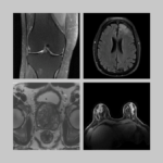
Raw k-space data and DICOM images from thousands of MRI scans of the knee, brain, and prostate, curated for machine learning research on image reconstruction.