
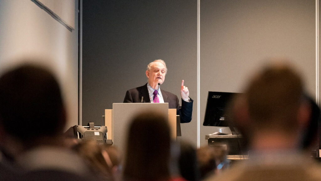
The Society for Cardiovascular Magnetic Resonance (SCMR) has conferred its 2023 gold medal award on Leon Axel, MD, PhD, professor of radiology, medicine, neuroscience and physiology at NYU Langone Health and scientist at the Center for Advanced Imaging Innovation and Research. The award, SCMR’s highest honor, was presented on January 27 in San Diego at the society’s annual meeting.
“Leon is honored for his contributions to the field of cardiac MR that go back many, many years,” said the society’s outgoing president Sven Plein, MD, PhD, as he announced the distinction. “He was the inventor of SPAMM, the [MRI] tagging that many of us still use today. He’s also trained numerous colleagues in cardiac MRI. Many of them went on to being presidents of this society.”
The technique referenced by Dr. Plein allows MRI to visualize the motion of the heart muscle by using radiofrequency pulses to “tag” the organ with a gridlike pattern. As the heart beats, the grid pattern deforms along with the tagged tissue, revealing in detail the heart’s contractions, relaxations, displacements, and distortions. Dr. Axel and his team created the method in the late 1980s and early 1990s at the University of Pennsylvania.
“The tagging, it was a way to really get a uniquely direct assessment of the regional motion inside the heart wall, which [otherwise] looks pretty featureless [on MRI] so it’s hard to really assess what’s going on,” said Dr. Axel. Although clinical implementation of the method and quantification of the heart’s motion proved to be challenging at the time, the idea became influential and inspired an entire subfield known as cardiac strain imaging.
“The whole concept of measuring regional [heart] function with MRI came from his lab,” said Karen Ordovas, MD, the incoming president of SCMR and professor of radiology at the University of Washington, where she is also section chief of cardiothoracic imaging. The tagging principle, developed further by other scientists, “evolved into what we use today which is called feature tracking and phase contrast measurements of strain,” she said.
Dr. Axel was also one of the founders of SCMR, established more than 30 years ago, in which he went on to serve in various roles, including as annual meeting organizer, secretary treasurer, committee and board member. “When we started, it was probably a couple of hundred people,” he said. “The question was do we need another society?” Today, SCMR’s membership stands at more than 5,000.
“Our main mission is to make sure that patients benefit from this technique—cardiac MRI—and have access to it,” said Dr. Ordovas. “The core of our membership is the U.S. and Europe, but we want to extend our impact beyond those regions.” She also sees more opportunity to expand access domestically. “In the United States, the technique is not fully adopted in some clinical applications … whereas in Europe it’s the bread and butter, especially in the U.K. and Germany,” she said. “There’s a lot of work for us to do.”
Before he pursued medical training, Dr. Axel had earned a doctorate in astrophysics (he described an atmospheric haze on Jupiter, later also identified on Saturn, known today as Axel dust). The desire to do something more people-oriented drew him to medicine, and in 1982—after completing medical school, residency, and fellowship at the University of California at San Francisco—he joined radiology faculty at the University of Pennsylvania, just as the arrival of MRI and CT scanners was setting off profound changes in the field. Armed with a physicist’s knack for studying basic phenomena and familiar with image processing principles pioneered in astronomy—such as the use of the Fourier transform to create pictures from electromagnetic signal—Dr. Axel found himself in the vanguard of advances that would shape medical imaging.
“In a lot of these things, I wasn’t the first person … but I was one of the first to try to help develop things,” Dr. Axel said. “For example, I was one of the first to use surface coils,” which he would also construct himself. Surface coils are now routine in imaging, as they increase the quality of data acquired from the anatomy of interest and underpin fast imaging techniques such as parallel MRI. “I’ve been involved with trying to develop ways of imaging blood flow in the heart … looking at perfusion imaging … and some early work in perfusion imaging in the brain with CT methods,” Dr. Axel said.
“He’s really considered a luminary in our field,” said Dr. Ordovas. “He has had a tremendous impact in education and mentoring, and his research ideas and developments have evolved over the years to impact patients with heart failure all over the world.” She added that nomination letters equally emphasized Dr. Axel’s achievements, mentorship, and character. “Leon is the kindest person you will ever meet,” she said.

Dr. Axel’s interest in the heart has not waned over the decades—far from it. He has continued to lead and participate in collaborative research to advance cardiac imaging with new technologies, including sparsity-based MRI reconstruction, extra-dimensional radial MRI, diffusion MRI, sodium MRI, and machine learning models. The heart, it turns out, still holds many mysteries.
“Surprisingly, although we can see many things visually even without the tagging, what we report in our official clinical MRI studies of the heart is an extremely limited set of data,” said Dr. Axel. “Basically, how much blood does the heart squeeze out with each beat—that’s it.” More detailed metrics of the heart’s function have the potential to uncover discrete pathology causes currently obscured by similar-appearing symptoms and lead to more effective therapies, explained Dr. Axel, who splits his time between conducting research and reading clinical cases.
The SCMR gold medal award “is a recognition that I played a role in the society and in the field,” said Dr. Axel. “We still have many potential things we can develop to make imaging even more clinically useful.” One of his current projects involves creating machine learning algorithms to characterize cardiac dyssynchrony with MRI. “It’s an ongoing story,” he said.