In a special issue of the journal Sensors dedicated to MRI, scientists with the Center for Advanced Imaging Innovation and Research at NYU Langone Health are sharing early experiences with alternative sensors for tracking motion in low-field MRI systems.
Anyone who has had an MRI can likely recall being told not to move in the scanner, but even when patients try to lie still, their bodies brim with motion. Much of this motion is caused by breathing, which shifts the shape and position of everything inside the abdomen—from the organs pressed up against the diaphragm, such as the the liver, down to structures resting on the pelvic floor, such as the bladder. In MRI, even subtle movement can result in artifacts, loss of definition, and less accurate measurements. Hence, medical imaging researchers have developed a multitude of techniques to track and correct for motion.
Now, a growing trend toward accessible low-field MRI—which has lower signal and higher noise than do standard clinical scanners—is stoking interest in supplemental sensors. At the same time, advances in electronics and machine learning are prompting scientists to imagine accessorizing MRI to make its data and images more accurate and more informative.
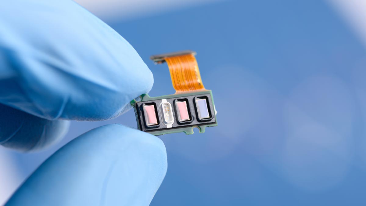
Exploring these new avenues, researchers at NYU Langone’s Center for Advanced Imaging Innovation and Research report early experiences at 0.55 Tesla with three alternative motion-sensing technologies: a time-of-flight camera, a pilot tone radio frequency transmitter, and an ultrasound transducer. They find that these systems offer complementary and partially overlapping information about motion at different depths: from the body’s surface to the major organs close beneath to the organs situated deep within the abdomen.
In a first, the research team has also tested the sensors apart from an MRI scanner. A setup independent of an MRI has “potential value for motion tracking during position-sensitive treatments such as radiation therapy,” write the authors.
Discussing their findings, the researchers envision a “sensor suite” that capitalizes on the advantages of each sensing mode and has the potential to “disentangle different motions that occur simultaneously … but are not related.” Such a suite, the authors write, “would enable the user to select between the different sensors depending on their strengths and capabilities based on the use case.”
A version of this post first appeared on the CAI2R LinkedIn.
Related Publication
Preliminary Experience with Three Alternative Motion Sensors for 0.55 Tesla MR Imaging.
Sensors (Basel). 2024 Jun 7;24(12):3710. doi: 10.3390/s24123710
Related Resource

A compact, stand-alone device for tracking and correcting patient motion in MRI exams.
Related Story

Imaging scientists around the world are looking into this simple transmitter to deal with breathing motion in MRI scans. NYU Langone offers resources to kickstart their research.