Eric Sigmund, PhD, is a professor in the department of radiology at NYU Grossman School of Medicine and an imaging scientist with the Center for Biomedical Imaging and the Center for Advanced Imaging Innovation and Research at NYU Langone Health. He investigates and develops a range of diffusion MRI methods, with particular focus on diffusion-weighted imaging (DWI), diffusion tensor imaging (DTI), and intravoxel incoherent motion (IVIM). He has explored novel applications in multiple organ systems and led NIH-supported research on new methods for muscle, breast, and kidney imaging, making substantial contributions to IVIM techniques for characterization of breast cancer, renal malignancy, and renal function. An authority in the growing area of IVIM imaging, Dr. Sigmund is a lead author of recent consensus recommendations for IVIM MRI. Our conversation was edited for clarity and length.
How would you describe the current focus of your research?
I work on diffusion MR techniques that collect, analyze, and interpret diffusion data in various clinical applications—a big one at the moment is oncology. We have ongoing breast cancer and renal cancer studies where water diffusion is sensitive to several key features of malignancy. Almost all tumors entail aggressive growth of cells, and that cellularity limits diffusion. They also often have abnormally elevated blood supply to feed that growth—that’s enhanced water motion, different from the Brownian thermodynamic motion of molecular water. We can sense and separate and quantify each of these. That’s one of the methods I work a lot with in my current research.
These techniques are also important in whole-organ contexts. The kidney, for example, receives a tremendous amount of perfusion to clean our blood supply and produce urine, and the diffusion analysis has to take that into account.
Can you talk about the techniques and imaging approaches that allow for the simultaneous probing of flow and diffusion? These are both types of fluid motion but they occur on very different scales.
The key is that within diffusion MR sequences, there’s a flexibility in how much you encode the contrast. When we encode what we call diffusion weighting, we are imposing an additional magnetic gradient beyond the imaging ones, and as the spins move, some move to the left, some move the right, they acquire phase different to one another, so they don’t maintain a coherence. That is the measurement, because that decoherence attenuates the overall signal. You can apply those gradients in many combinations, and you can change their strength, separation, direction. What you measure is the overall displacement—whether the thermodynamic one [diffusion] or the driven one [flow]—and the overall property that describes what the spins are doing at an ensemble level can be a multidimensional object with multiple contributions.
The perfusion process is not Brownian, but it is dispersive and it still amounts to incoherent motion—in the literature it’s called a pseudo-diffusion process. The way you separate that is by incrementally encoding the strength of a gradient in a set of measurements so that you’re not just measuring one decay rate but can resolve multiple decay rates, multiple populations within the system. These can also be brought out with different types of diffusion encoding, or timing within the heartbeat cycle. There are different numerical ways to process the data to make sure that that decomposition is good. Depending on the organ and what signals are present, you can often read out more than one flow component: the very fast, the intermediate, and what remains.
And looking at the different ‘populations’ allows you to measure or estimate different properties of flow and diffusion within a given voxel?
That’s the idea. In the kidney example, you have a vascular system and a tubular system: two types of flow but at different rates. Vascular flow is typically faster than is tubular flow. If you read out those two speeds separately, then they’re assumed to originate from those two sub-volumes of the organ. As with all diffusion imaging, it’s a combination of assumptions, modeling, and some acknowledgment that you’re reading out what is emergent from the entire ensemble as an average, rather than tracking every single spin, which is unattainable.
I have a Galton board on my desk—it’s a good illustration of the Gaussian distribution. What you have here is a one-dimensional diffusion process: every time a pebble hits a pin it can go left or right, and after hitting a number of pins it ends up in a chamber on the bottom. The most common result is to go nowhere, and that’s the peak of the distribution, but there’s also the probability to go elsewhere. The idea is that even though you don’t know what each individual pebble is going to do, you know exactly what the distribution will be.
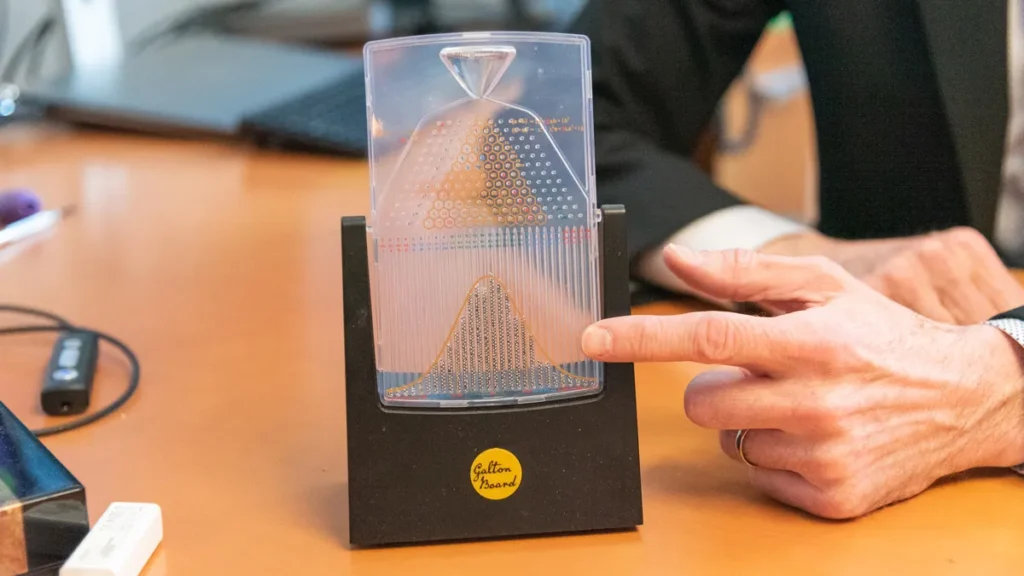
It’s a lesson in both physics and statistics.
It can be equally appropriate on a physicist’s desk and an economist’s desk. Here, it illustrates the principle that diffusion properties are ensemble properties.
The separation of structure and flow is typically called intravoxel incoherent motion, or IVIM. Another technique called diffusion tensor imaging—DTI—focuses on directionality. When the primary variable is the direction of the diffusion and it’s larger in one direction than another, it shows that local microstructure has a preferred orientation. There are ways to use IVIM and DTI in concert because flow is also directional. Again, an example in the kidney: in the medulla—the pyramid where you have a parallel set of tubules where some of the filtration takes place—there is active flow. That is a case in which a hybrid of these approaches is needed to describe the kidney in the best way.
Different types of motion happening in multiple directions at different scales make the kidney sound like a challenging environment for magnetic resonance techniques.
What I like about that is that within the world of diffusion everyone can find a zone where they can innovate. Here, it’s a combination of microstructure and microfluidics. It’s an opportunity to see new features, explore them, and learn how they connect with pathologies.
When you brought up cancer earlier, you said that in tumors diffusion tends to be restricted but perfusion tends to be increased. That sounds a bit counterintuitive.
That’s right—in the case of cancer tissue, they are competing influences, so using a single-component model would be limiting. That’s another reason to do the type of imaging we do: to not confuse those features. What resolves that counterintuitive thought is that these features originate from different spaces: one from the vascular system, the other from the parenchyma, or cellular bed. Different spins are telling you about the vascularity and the cellularity, and these two features are often quite important in aggressive tumors.
And various combinations of diffusion and flow parameters give IVIM techniques a wide array of potential uses as proxies for pathology?
The more you measure the more you can differentiate—either among types of tumors or between tumors and native tissue or how tumors respond to therapy.
The community that does IVIM had a workshop a couple of years ago—to catch up on the latest research but also to come up with recommendations about how to do the measurements going forward in as consensus-driven a way as possible, so that we could pool evidence when it comes time to add things to clinical trials and consider seeking FDA approval for certain indications, because that comes with steep requirements for n number and effect size.
That effort led to a consensus paper, or white paper. We reviewed the literature to date and all the meta-analyses reported in different organs, and one plot we generated shows the spectrum of those two components—diffusion and perfusion fraction values—across at least six different organ systems. It’s quite a constellation, because each organ has its own signature. That goes to show the progression of the field. IVIM is no longer in a pilot stage and is hopefully entering a translational phase.
It’s one thing to conduct experiments in your own lab according to whatever criteria you think best serve your setup, your hardware, your pre- and post-processing pipelines and your analysis, and another thing to try to define standards for everyone. Can you say more about that?
We each have a need and interest in innovating on one’s own, and I like to think of it as akin to being a violin soloist versus playing in an orchestra. If we all try to be soloists all of the time, that will limit our progress. We have an ongoing study focusing on broadening translation of IVIM for the specific context of breast cancer, for example, involving analysis of patient data from five worldwide sites. Sometimes we have to build an evidence level with a certain agreed-upon version of a technique. Going deeper and going broader are not necessarily mutually exclusive.
There are different perspectives on how to translate IVIM next. How much should be done in precisely the same way? How much flexibility does one want to have? How much should we recommend that clinicians take IVIM into account, and when? And what do you want to target: changing day-to-day clinical practice or changing a clinical trial that depends on the imaging to evaluate a particular therapy? There’s no perfect answer because everyone has different interests.
How do the recommendations accommodate both the solo innovation and the ensemble science?
There are tiers of suggested protocols: an abbreviated level of measurements we can all agree on, so that we can compare the numbers from different sites; an intermediate minimal tier, if you want to collect one more parameter—again, in a way that standardizes the encoding; and then an advanced level, which is anything else you’d like to add. The advanced strategy retains the encodings that are also in the abbreviated and minimal strategy, so that in the future one can peel out the common parts of the protocol and build up that evidence base. The philosophy is don’t limit your own research but try to create a common thread that can contribute to generalized evidence.
In other words, be a good citizen of the scientific community.
Or at least allow the data to exist so that someone can be.
One of the areas that you have innovated in is dynamic imaging of skeletal muscle. Can you talk a little bit about that part of your work?
That’s a longer story, so let me give just a bit of a background on my trajectory to medical imaging. I did my doctorate in condensed-matter physics in a nuclear magnetic resonance lab. In my thesis I focused on glass-forming liquids and very slow motion. At that frontier, the stronger the gradients you could apply, the slower the diffusion you could sense. And one of the strongest gradients is not the one applied by a coil with a lot of current but the one at the edge of a superconducting magnet—it’s called a fringe field gradient.
Like a natural, static gradient that’s just there.
That’s right, so we would arrange for the sample to be in that position, cool the systems down, and watch the diffusion slow to a very low level to understand the thermodynamics or the ion transport of the different parts of an electrolyte. That research taught me gradient measurements and along the way I became interested in MRI as a branch of MR. That interest led me do a postdoc in clinical MRI at Northwestern Hospital in Chicago and later to a stint in an industrial lab at Schlumberger [now known as SLB] applying rock techniques to trabecular bone. I was also contributing to ongoing research on a method for collecting complex diffusion information in the ground while searching for oil. The idea was to get many echoes and vary the acquisition to move the direction of the gradients in order to collect the directionality information quickly.
Working on that, I was already aware of medical applications like diffusion MRI in the brain, where you have the directionality of white-matter fibers. But this method was much faster, and when I started at NYU Langone in 2006, one of my goals was to see whether it could be converted for use on a clinical scanner.
Instead of collecting directional information one shot at a time, the concept was to get all those directions in a single shot, so that if you wanted to capture some dynamic process, you could. Making it work on a clinical scanner in real tissue was quite an engineering feat. My first postdoc—Steven Baete, who is now faculty at NYU Langone—did that, redesigning the pulse structure and the k-space acquisition to allow for motion correction in order to collect data in a dynamic way but with a full DTI information set.
The target—coming around to the question—was to collect DTI dynamically in skeletal muscle following exercise. Because the act of exertion, of contracting our muscles, calls for perfusion: it involves fluid exchange, changes in the microstructure of the muscle fibers, change in water content surrounding those fibers—and that’s all transient.
In other words, a single-shot diffusion tensor imaging approach allowed you to measure more frequently in an environment where lots of things are changing fast.
I have to admit, it was a hammer looking for a nail, and dynamic DTI in muscle was our contribution there. We ultimately also paired it with commercial ergometers which allowed exercise in the scanner for calf muscles and thigh muscles, to control the exertion and measure right away.
What was it like for you to transition from working with materials to working with biology, physiology, and medicine?
A lot of people in the MRI research community have come from different backgrounds: some from a very medical side, some from a very technical side, some starting from a hardware side. What I found is that the field—both here at NYU Langone and out there more broadly—is very collegial.
Our primary imaging community, the International Society for Magnetic Resonance in Medicine, was welcoming to me despite the fact that I was coming from a particular lane—very good on some things but not very educated on other things. I would be able to go to an ISMRM meeting and buttonhole people who were leaders of the field, and they’d be happy to have long conversations despite knowing even more about how much I didn’t know than I myself did at that point.
You have some interesting objects in your office—you brought up the Galton board earlier, and there’s also a cotton candy machine. Can you explain why it’s here?
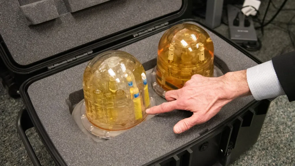
A lot of the times you want to have phantoms—synthetic standards that mimic the physiological processes that we want to measure. In the case of incoherent motion, vascular or tubular flow amounts to flow through a random network of tubes, and we’ve had models that simulate incoherent motion embedded in standard breast phantoms, for example, using a cellulose sponge, which redirects water flow in incoherent ways and generates a reliable, even if not entirely physiological, distribution of velocities.
One step toward something more biomimetic was noticed by a group led by Leon Bellan, who were looking for a way to mimic the vascular system to study how cells grow when fed by a certain amount of perfusion. They found that cotton candy makes a very close analog to a vascular network and came up with a recipe to make cotton candy mesh, embed it in something solid, and then dissolve it. What you’re left with is the negative space of the cotton candy, which gives you a bunch of channels that if you can flow through, you’ve simulated capillary flow. It’s called a sacrificial sucrose network, and it’s been taken up by MRI groups who use it to create phantoms. We’d like to do the same thing here to test our techniques.