
Ruoxun Zi has successfully defended her PhD thesis in biomedical imaging and technology at NYU Grossman School of Medicine. The defense was held on Wednesday, April 30, at NYU Langone Health.
In her doctoral research, Dr. Zi explored and expanded the current limits of radial sampling methods and iterative image reconstruction techniques by developing new applications in low-field MRI, neuroimaging, body imaging, and musculoskeletal imaging. Her dissertation was co-advised by Tobias Block, PhD, associate professor of radiology at NYU Langone; and Riccardo Lattanzi, PhD, professor in the department of radiology at NYU Langone and director of NYU Langone’s Center for Biomedical Imaging (both Dr. Block and Dr. Lattanzi are scientists with the Center for Advanced Imaging Innovation and Research).
Radial sampling is a way of acquiring magnetic resonance imaging data in “spokes” or “blades” that fill MRI’s raw-data dimension in an asterisk-like pattern, a technical feature sometimes reflected in the names of radial pulse sequences, such as Stack of Stars or PROPELLER. Imaging methods that rely on radial sampling have greater computational complexity than the traditional Cartesian acquisitions, where data fill an array row-by-row, but come with a number of benefits in three important areas: acceleration, post-processing flexibility, and compatibility with patient motion.
In one project, reported in 2024 in the journal Magnetic Resonance in Medicine, Dr. Zi and colleagues developed a fat suppression method for low-field MRI. Fat suppression is important in MRI because the naturally bright appearance of adipose tissue on MR images may obscure lesions and make tissue types difficult to distinguish, somewhat analogous to how details may be blown out in overexposed photos. Low-field MRI, an area of growing scientific interest, has created the need to improve a range of techniques—including fat suppression—that have become rudimentary at the higher fields used in clinical imaging. Authors note that the new method is also applicable to high-field MRI with strong B0 field inhomogeneities and can potentially be extended to quantify other MRI metrics, such as proton density fat fraction.
Another investigation led by Dr. Zi focused on creating radial variants of an MRI sequence recently proposed for cancer imaging, shown to produce superior tumor-to-brain contrast and vascular detail than do current clinical counterparts. The approach is called T1 relaxation-enhanced steady-state (T1RESS). Dr. Zi and colleagues have developed radial T1RESS adaptations and tested them in phantoms, people diagnosed with brain cancer, and healthy controls. Detailing their findings in the journal Investigative Radiology, the researchers write that their sequences “offer improved lesion conspicuity and motion robustness and enable dynamic imaging for contrast-enhanced brain MRI.”
In yet another study, Dr. Zi explored combining a radial imaging method called GRASP, with a motion detection technique known as the Pilot Tone—both previously developed at the Center for Advanced Imaging Innovation and Research—to track patient motion in abdominal and pelvic MRI. The natural movement of internal organs, which shift and flex during breathing, poses challenges in MRI. Techniques like GRASP and the Pilot Tone are aimed at mitigating or tracking these shifts in order to obtain sharp images while allowing patients to breathe normally during scans. In her abstract, Dr. Zi writes that the process she developed for tracking motion information “resulted in improved image sharpness and temporal consistency.”
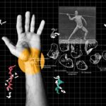
Last, Dr. Zi also led the development of an MRI method to examine and quantify the kinematics of natural wrist motion, an area that is not well understood. The project required Dr. Zi and colleagues to address the dual challenges of imaging in three dimensions and at a high temporal resolution. In order to quantify motion, the team created new imaging hardware, an image reconstruction pipeline, and image segmentation methods that make possible the estimation and comparison of the displacement of each of the wrist’s eight bones. The research was covered on this blog and published in the journal Skeletal Radiology, where the authors write that their work “demonstrates that real-time volumetric dynamic examination of the moving wrist is feasible” and could enable new kinds of MRI assessments for wrist instability.
Related Publications
Fat suppression using frequency-sweep RF saturation and iterative reconstruction.
Magn Reson Med. 2024 Nov;92(5):1995-2006. doi: 10.1002/mrm.30199
T1 Relaxation-Enhanced Steady-State Acquisition with Radial k-Space Sampling: A Novel Family of Pulse Sequences for Motion-Robust Volumetric T1-Weighted MRI with Improved Lesion Conspicuity.
Invest Radiol. 2025 Apr 7. doi: 10.1097/RLI.0000000000001185
High-resolution volumetric dynamic magnetic resonance imaging of the wrist using an 8-channel flexible receive coil.
Skeletal Radiol. 2025 Jun;54(6):1291-1299. doi: 10.1007/s00256-024-04829-7
Related Stories
Imaging researchers at NYU Langone Health are getting close to quantifying the dynamics of natural wrist motion, which are not well understood.

Imaging scientists around the world are looking into this simple transmitter to deal with breathing motion in MRI scans. NYU Langone offers resources to kickstart their research.